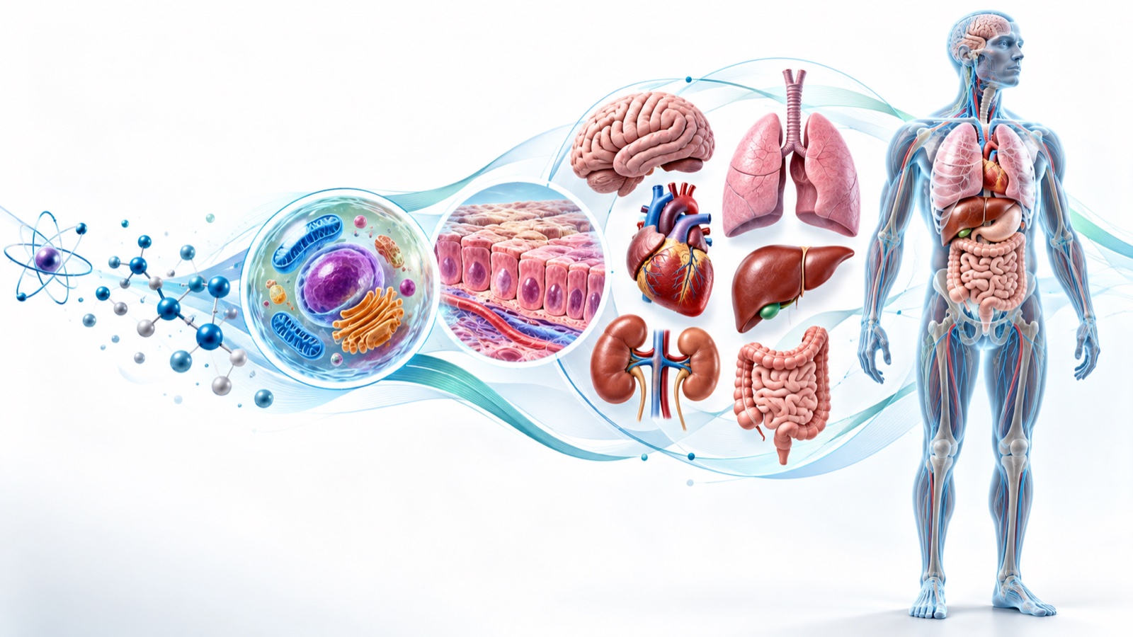
How Chemicals Cross Cell Membranes: The Foundation of Human Life and Homeostasis
An illustrated nursing guide to how oxygen, water, glucose, and electrolytes move in and out of cells, and how diffusion, osmosis, and active transport connect to homeostasis, IV fluids, and NCLEX reasoning.
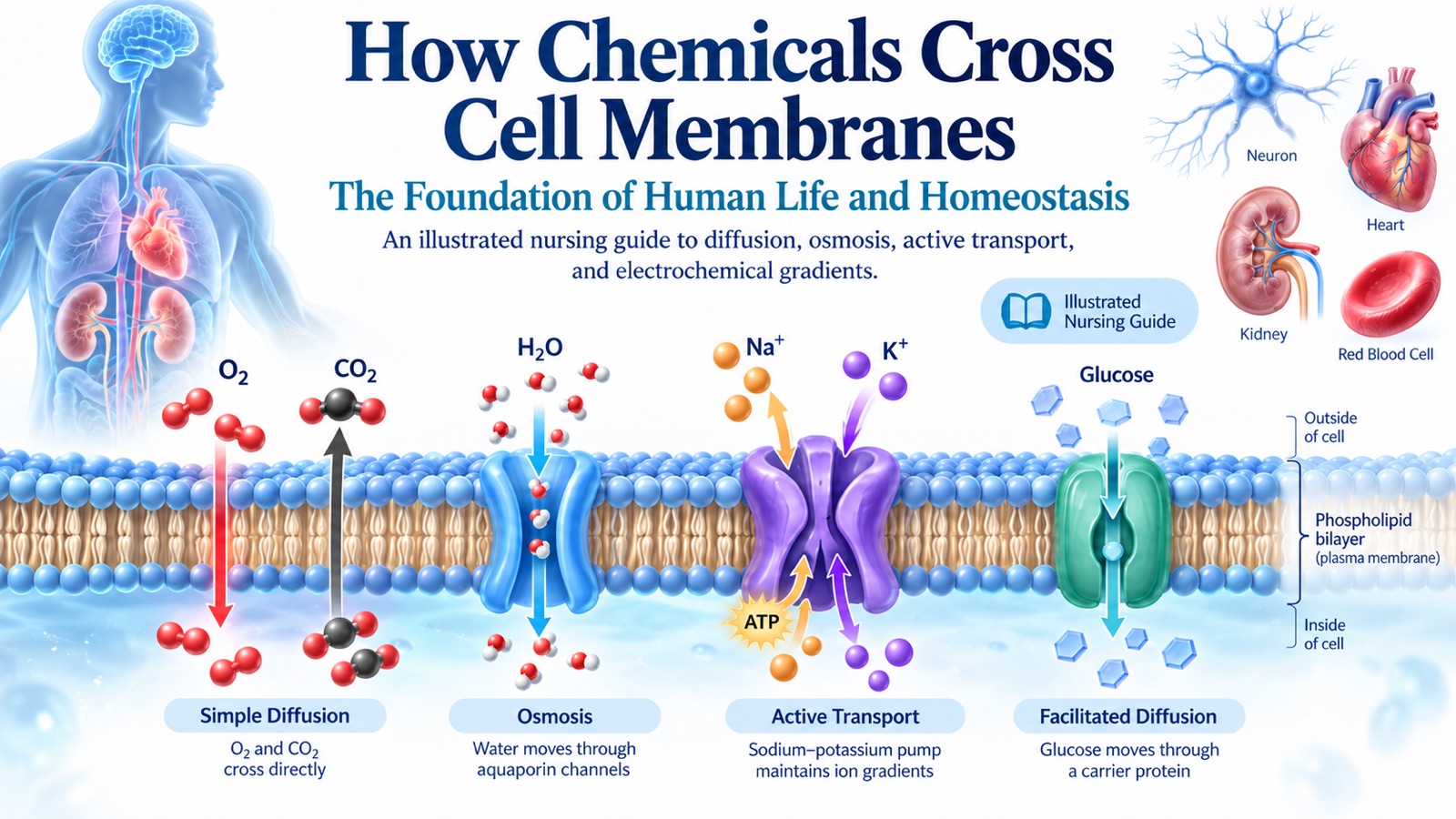
Every heartbeat, nerve impulse, muscle contraction, and cellular reaction depends on one quiet, constant process: substances moving across cell membranes.
In From Atoms to Organism: The 8 Levels of Human Body Organization, we saw how a single potassium ion can eventually affect the entire patient. This guide zooms in on the boundary where that story begins — the cell membrane — and explains how oxygen, water, glucose, electrolytes, and larger molecules get in and out of cells.
Along the way, we will connect each mechanism to homeostasis, common clinical problems, and the kind of reasoning the NCLEX expects.
Quick Overview
| Mechanism | Needs a Membrane Protein? | Uses Energy Directly? | Classic Example |
|---|---|---|---|
| Simple diffusion | No | No | O₂ entering cells, CO₂ leaving |
| Facilitated diffusion | Yes (channel or carrier) | No | Glucose entering muscle through GLUT4 |
| Osmosis | Often (aquaporins speed it up) | No | Red blood cell swelling in a hypotonic solution |
| Filtration | No (pressure-driven) | No | Fluid filtering out of the glomerulus |
| Primary active transport | Yes (pump) | Yes (ATP) | Na⁺/K⁺ pump: 3 Na⁺ out, 2 K⁺ in |
| Secondary active transport | Yes (cotransporter) | No — uses an existing Na⁺ gradient | SGLT1 absorbing sodium and glucose together |
| Vesicular transport | Membrane vesicles | Yes | White blood cells engulfing bacteria |
Why Cell Membrane Transport Matters
The human body contains trillions of cells, but no cell can survive by simply sealing itself off from its surroundings. A cell must obtain oxygen and nutrients, control its water and electrolyte content, communicate with other cells, and manage metabolic byproducts.
These exchanges occur continuously through the plasma membrane, also known as the cell membrane.
Consider the different needs of a few specialized cells:
- Neurons maintain ion gradients to transmit electrical signals.
- Cardiac muscle cells regulate sodium, potassium, and calcium to coordinate heartbeats.
- Kidney tubular cells transport salt, water, glucose, and acid–base components to regulate the body's internal environment.
- Intestinal epithelial cells absorb nutrients using dedicated membrane proteins.
- Red blood cells exchange gases and regulate their volume as they pass through the circulation.
All these processes contribute to homeostasis, the maintenance of internal conditions within ranges compatible with normal function.
To understand homeostasis at the organ level, we must first understand transport at the cellular level.
1. The Cell Membrane: A Selective Barrier
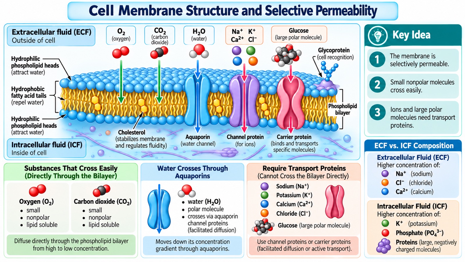
Figure 1. The plasma membrane separates intracellular and extracellular fluid while controlling the passage of different substances.
1.1 The Structure of the Plasma Membrane
The plasma membrane is primarily a phospholipid bilayer.
Each phospholipid has two distinct components:
- Hydrophilic head: Attracted to water and faces the watery environment inside or outside the cell.
- Hydrophobic fatty acid tails: Repel water and face inward toward each other.
This arrangement creates a barrier between the cell's internal environment and its surroundings.
Small, nonpolar molecules such as oxygen (O₂) and carbon dioxide (CO₂) can cross this barrier relatively easily.
However, charged ions such as sodium (Na⁺), potassium (K⁺), and calcium (Ca²⁺) cannot efficiently cross the lipid interior without specialized membrane proteins.
Glucose is also too polar to cross efficiently without dedicated transporters.
1.2 Important Membrane Components
The plasma membrane contains several other components:
- Channel proteins: Provide selective pathways for ions and water.
- Carrier proteins: Bind specific substances and change shape to move them across the membrane.
- Cholesterol: Influences membrane stability and fluidity.
- Glycoproteins: Participate in cell recognition, communication, and signaling.
- Glycolipids: Contribute to membrane recognition and interactions.
These structures help determine which substances enter or exit cells and how rapidly they move.
1.3 Intracellular Fluid vs. Extracellular Fluid
The body's fluid is divided into two major compartments.
Intracellular fluid (ICF) is the fluid inside cells.
Extracellular fluid (ECF) is the fluid outside cells, including interstitial fluid and blood plasma.
Their chemical compositions differ significantly.
| Substance | Relatively More Abundant | Primary Importance |
|---|---|---|
| Sodium (Na⁺) | Extracellular fluid | Fluid regulation, electrical signaling |
| Chloride (Cl⁻) | Extracellular fluid | Electrical balance, fluid balance |
| Ionized calcium (Ca²⁺) | Extracellular fluid | Muscle contraction, cellular signaling |
| Potassium (K⁺) | Intracellular fluid | Membrane potential, cellular excitability |
| Phosphate | Intracellular fluid | ATP production and cellular metabolism |
| Many proteins | Intracellular fluid | Metabolism, cell structure, buffering |
Key Concept: Cells remain alive not because their internal environment is identical to their surroundings, but because they continuously maintain important chemical differences.
2. What Substances Enter and Exit Human Cells?
Different chemical substances serve different biological purposes.
Understanding their functions makes membrane transport much easier to learn.
2.1 Essential Substances
| Category | Examples | Major Function |
|---|---|---|
| Respiratory gases | Oxygen, carbon dioxide | Aerobic metabolism and CO₂ elimination |
| Water | H₂O | Cell volume, fluid balance, chemical reactions |
| Electrolytes | Na⁺, K⁺, Ca²⁺, Mg²⁺, Cl⁻ | Nerve conduction, muscle contraction, electrical balance |
| Nutrients | Glucose, amino acids, fatty acids | Energy production, tissue repair, biosynthesis |
| Acid–base substances | H⁺, HCO₃⁻ | Regulation of cellular and extracellular pH |
| Signaling molecules | Neurotransmitters, hormones | Cellular communication |
| Metabolic products | CO₂, lactate, urea | Transport, reuse, or eventual elimination |
2.2 Important Electrolyte Reference Ranges
| Electrolyte | Typical Adult Serum Reference Range |
|---|---|
| Sodium | 135–145 mEq/L |
| Potassium | 3.5–5.0 mEq/L |
| Chloride | 98–106 mEq/L |
| Total Calcium | Approximately 8.5–10.5 mg/dL |
| Magnesium | Approximately 1.7–2.2 mg/dL |
| Bicarbonate | Approximately 22–28 mEq/L |
| Phosphorus | Approximately 2.5–4.5 mg/dL |
Note: Reference ranges vary by laboratory, patient population, and testing method.
An important nursing distinction is that a serum electrolyte measurement does not necessarily represent the body's total electrolyte stores.
For example, most potassium is stored inside cells.
Consequently, the serum potassium concentration represents only a small portion of the body's total potassium.
Nevertheless, changes in serum potassium can dramatically affect cardiac and neuromuscular function.
3. What Causes Substances to Move Across Membranes?
Substances move because physical and chemical forces act upon them.
These forces are known as gradients.
3.1 Concentration Gradient
A concentration gradient is a difference in the concentration of a substance between two regions.
For an uncharged substance, passive diffusion generally produces net movement from higher concentration toward lower concentration.
For example, oxygen can move from extracellular fluid into cells when oxygen partial pressure is higher outside the cell.
3.2 Electrical Gradient
An electrical gradient occurs when electrical potential differs between two regions.
Because ions carry electrical charges, their movement is influenced by membrane voltage.
For example, positively charged sodium ions are attracted toward a relatively negative cellular interior.
3.3 Electrochemical Gradient
The electrochemical gradient combines:
- The chemical effect of a concentration difference.
- The electrical effect of membrane voltage.
An ion's net movement through an open channel depends on the combined influence of these forces.
This explains why the direction of ion transport cannot always be predicted from concentration alone.
3.4 Osmotic and Pressure Gradients
Water movement is influenced by differences in water chemical potential, often explained in nursing as osmotic differences involving effectively nonpenetrating solutes.
Bulk movement of fluid can also occur in response to hydrostatic pressure differences.
These principles are essential for understanding fluid balance, edema, and kidney function.
4. Passive Transport: Movement Without Direct ATP Consumption
Passive transport occurs when substances move down their relevant gradients without direct ATP expenditure by the transport process.
Major examples include:
- Simple diffusion.
- Facilitated diffusion.
- Osmosis.
Filtration is another important pressure-driven transport process.
4.1 Simple Diffusion
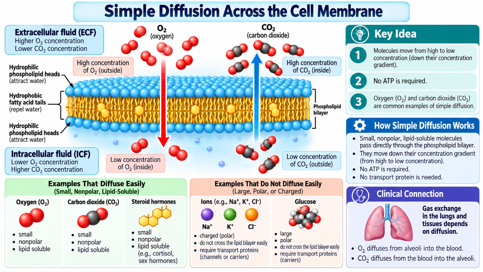
Figure 2. Oxygen and carbon dioxide cross the cell membrane through simple diffusion.
Simple diffusion is the net movement of molecules down their chemical potential gradient without requiring membrane transport proteins or direct ATP expenditure.
Oxygen normally moves from regions of higher oxygen partial pressure to regions of lower oxygen partial pressure.
Cells continually consume oxygen during aerobic metabolism, helping maintain conditions that favor oxygen diffusion into the cell.
Carbon dioxide is produced during metabolism and commonly diffuses outward into the surrounding fluid and eventually the blood.
Although individual molecules move randomly in both directions, their net movement follows the gradient.
Factors Affecting Diffusion
The rate of diffusion depends on several factors:
- Concentration or partial-pressure difference.
- Membrane surface area.
- Membrane thickness.
- Permeability of the membrane.
- Properties of the diffusing molecule.
Clinical Nursing Connection
Oxygen and carbon dioxide exchange in the lungs depends heavily on diffusion.
Oxygen diffuses from the alveoli into pulmonary capillary blood, while carbon dioxide diffuses in the opposite direction.
Disorders that impair gas exchange can reduce oxygen delivery to tissues.
However, adequate tissue oxygenation also depends on circulation, hemoglobin, and cellular oxygen utilization.
4.2 Facilitated Diffusion
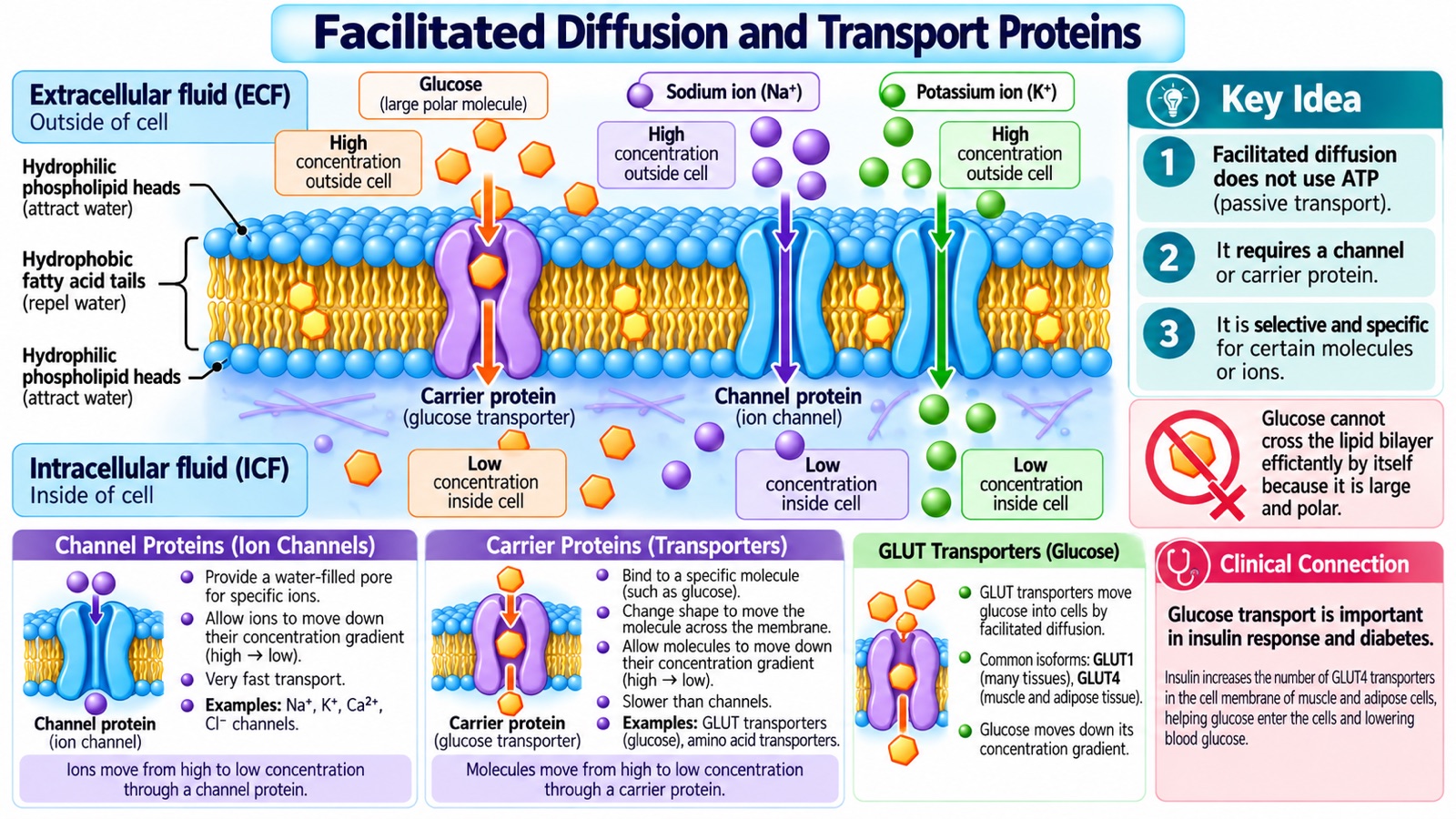
Figure 3. Membrane proteins allow specific molecules and ions to cross membranes down their gradients.
Facilitated diffusion occurs when molecules or ions move through membrane proteins without direct ATP consumption.
Unlike simple diffusion, facilitated diffusion requires a specialized transport protein.
There are two major types.
A. Channel Proteins
Channel proteins create selective pathways through the membrane.
Examples include:
- Sodium channels.
- Potassium channels.
- Calcium channels.
- Chloride channels.
- Water channels, called aquaporins.
Many ion channels are regulated by voltage changes, chemical signals, or mechanical forces.
When a channel is open, ions move according to their electrochemical gradients.
B. Carrier Proteins
Carrier proteins bind specific molecules and undergo conformational changes that transport those molecules across the membrane.
Carrier-mediated transport is often saturable because the number and activity of transporters are limited.
Example: Glucose Transport
Glucose is an important energy source, but it cannot efficiently cross the hydrophobic lipid bilayer by itself.
Instead, many cells rely on glucose transporters called GLUT proteins.
Examples include:
- GLUT1: Important in numerous tissues and basal glucose uptake.
- GLUT4: Important in skeletal muscle and adipose tissue.
Insulin signaling increases the number of GLUT4 transporters available at the cell surface.
This allows increased glucose uptake into insulin-responsive tissues.
Clinical Nursing Connection
Glucose transport is closely connected to:
- Diabetes mellitus.
- Insulin physiology.
- Blood glucose regulation.
- Exercise.
- Cellular energy metabolism.
Understanding facilitated diffusion helps explain how insulin promotes glucose uptake and why insulin resistance affects metabolism.
4.3 Osmosis and Tonicity
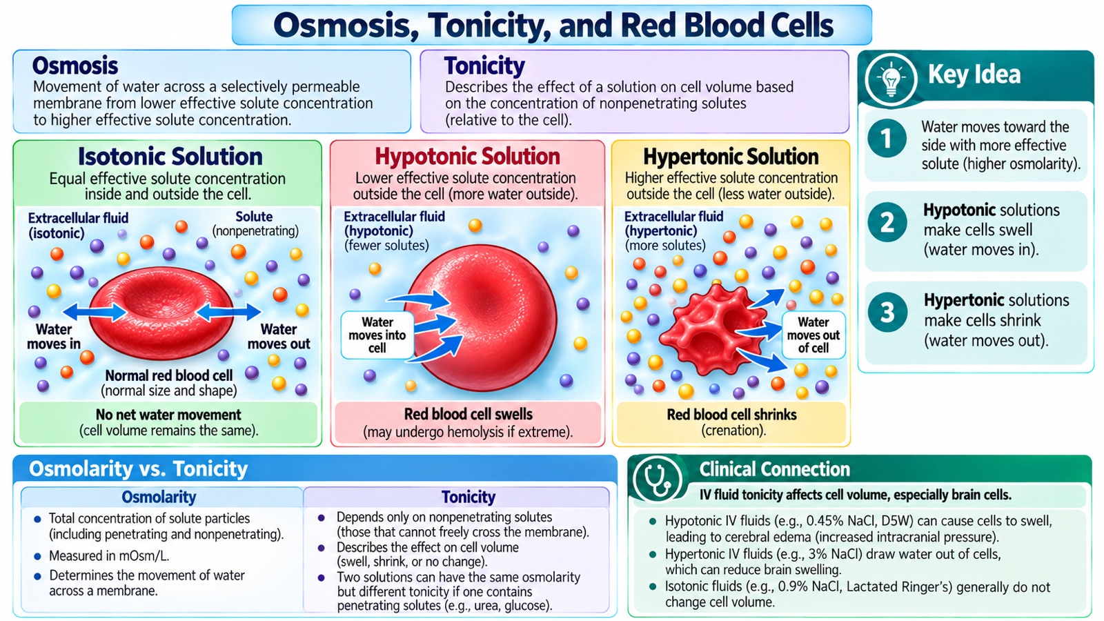
Figure 4. Changes in extracellular tonicity affect the direction of water movement and cell volume.
Osmosis describes the movement of water across a selectively permeable membrane in response to differences in water chemical potential.
For introductory nursing applications, water tends to move toward the side with a higher concentration of effectively nonpenetrating solutes.
Water can cross membranes directly to some extent, but specialized channels called aquaporins allow rapid water movement in many tissues.
A. Isotonic Solution
In an isotonic solution, effective osmotic forces inside and outside a cell are balanced.
Water moves in both directions without sustained net movement.
Result: The cell maintains approximately normal volume.
B. Hypotonic Solution
A hypotonic solution has a lower concentration of effectively nonpenetrating solutes than the intracellular environment.
Water moves into the cell.
Result: The cell swells.
Extreme swelling may cause cell rupture, known as lysis.
In red blood cells, this is called hemolysis.
C. Hypertonic Solution
A hypertonic solution has a higher concentration of effectively nonpenetrating solutes than the intracellular environment.
Water moves out of the cell.
Result: The cell shrinks.
In red blood cells, this shrinking can produce a crenated appearance.
Osmolarity vs. Tonicity
Although these terms are related, they are not identical.
Osmolarity measures the total concentration of osmotically active particles per liter of solution.
Tonicity describes the effect of a solution on cell volume, largely determined by solutes that cannot readily cross the membrane.
For example, urea contributes to osmolarity but is relatively ineffective at maintaining tonicity across many cell membranes.
Clinical Nursing Connection: IV Fluids
Understanding tonicity is essential when administering intravenous fluids.
Examples include:
- 0.9% Sodium Chloride: Commonly considered an isotonic crystalloid.
- Lactated Ringer's: A balanced crystalloid usually grouped with isotonic fluids in nursing practice.
- 0.45% Sodium Chloride: Hypotonic relative to normal plasma.
- 3% Sodium Chloride: Hypertonic saline.
- D5W: Approximately isotonic in the container, but produces a physiologically hypotonic effect after glucose metabolism.
Fluid selection depends on the patient's condition, sodium concentration, volume status, and treatment goals.
Improper administration can contribute to dangerous fluid shifts.
4.4 Filtration
Filtration is movement of water and small solutes driven by pressure differences.
One of the most important examples occurs in the kidneys.
Inside the glomerulus, blood pressure contributes to filtration across a specialized capillary barrier.
Water and many small solutes enter the nephron, while blood cells and most large proteins remain in the circulation.
This process is fundamental to urine formation.
Clinical Nursing Connection: Changes in kidney filtration can affect fluid balance, electrolyte regulation, and waste elimination.
5. Active Transport: How Cells Maintain Chemical Gradients
Cells cannot survive by relying on passive transport alone.
Many important ions must be maintained at different concentrations inside and outside cells.
Active transport helps preserve these differences.
5.1 Primary Active Transport: The Sodium–Potassium Pump
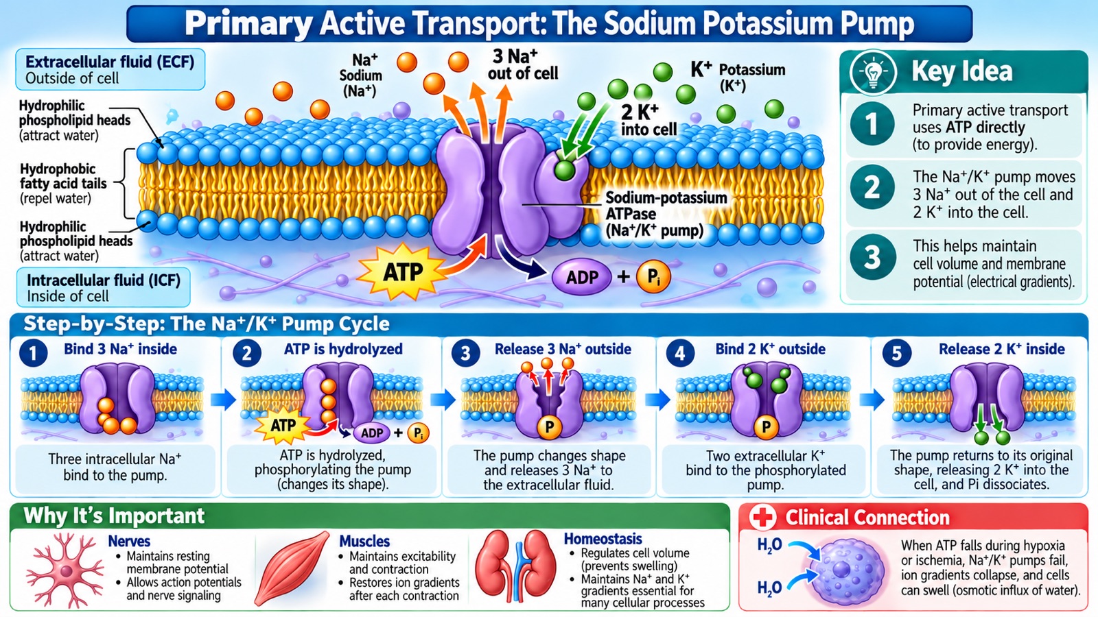
Figure 5. The sodium–potassium ATPase uses ATP to maintain sodium and potassium gradients.
Primary active transport directly uses energy, often from ATP hydrolysis, to move substances against their electrochemical gradients.
An essential example is the sodium–potassium ATPase (Na⁺/K⁺ pump).
During a typical pump cycle:
- Three Na⁺ ions bind from inside the cell.
- One ATP molecule is hydrolyzed.
- The pump undergoes conformational changes and releases three Na⁺ ions outside.
- Two K⁺ ions bind from outside.
- The pump returns toward its initial conformation and releases two K⁺ ions inside.
Therefore:
3 Na⁺ out + 2 K⁺ in + 1 ATP hydrolyzed
The pump helps maintain:
- High extracellular sodium concentration.
- High intracellular potassium concentration.
- Cellular volume regulation.
- Normal membrane excitability.
- Gradients required for secondary active transport.
The pump is also electrogenic because it exports more positive charge than it imports.
What Happens When ATP Becomes Unavailable?
Consider a cell experiencing severe oxygen deprivation.
If ATP production becomes inadequate:
- ATP-dependent membrane pumps become less effective.
- Sodium and other ionic gradients begin to deteriorate.
- Intracellular ion balance becomes abnormal.
- Water may accumulate inside the cell.
- Cellular swelling develops.
- Prolonged severe injury can lead to irreversible damage or cell death.
This mechanism contributes to cytotoxic edema during ischemic injury.
Clinical Nursing Connection
Understanding the sodium–potassium pump helps explain:
- Cellular injury caused by ischemia.
- Cerebral edema.
- Nerve and muscle excitability.
- Cardiac electrophysiology.
- The importance of adequate ATP production.
5.2 Secondary Active Transport: Sodium–Glucose Cotransport
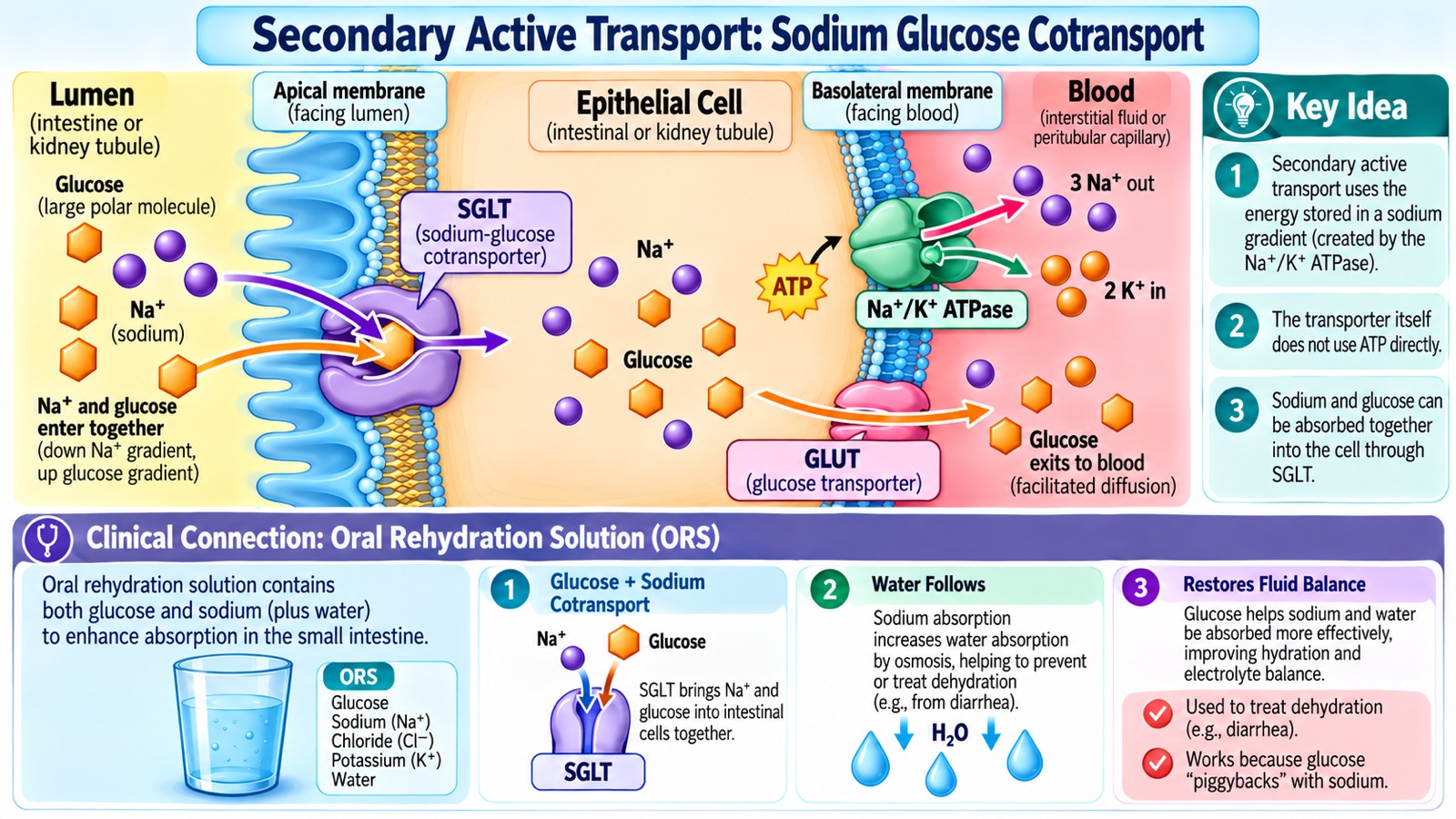
Figure 6. Sodium and glucose can enter intestinal epithelial cells together by secondary active transport.
Secondary active transport uses the energy stored in an existing electrochemical gradient.
The transporter itself does not directly hydrolyze ATP during each transport cycle.
Instead, it uses a gradient established by another energy-consuming process.
Example: Sodium–Glucose Cotransporter
In the small intestine, an important transporter is SGLT1.
SGLT1 uses the inward sodium gradient to transport glucose into intestinal epithelial cells.
The process works as follows:
- The basolateral Na⁺/K⁺ ATPase maintains a low intracellular sodium concentration.
- Sodium tends to enter the cell down its electrochemical gradient.
- SGLT1 couples sodium entry with glucose uptake.
- Glucose exits through a basolateral glucose transporter, primarily GLUT2.
- Sodium absorption contributes to osmotic water absorption.
This system allows glucose absorption even when the glucose concentration gradient alone would not favor its entry.
Clinical Nursing Connection: Oral Rehydration Therapy
Oral rehydration solutions contain glucose and electrolytes.
The presence of sodium and glucose supports intestinal absorption through SGLT1.
Water absorption accompanies solute transport.
This is particularly useful in treating dehydration associated with many diarrheal illnesses.
A related transporter, SGLT2, contributes to glucose reabsorption in the kidneys.
SGLT2 inhibitors are used in several clinical settings, including the management of certain patients with diabetes, heart failure, or chronic kidney disease.
5.3 Other Important Pumps and Exchangers
| Transport Protein | Primary Function | Clinical Connection |
|---|---|---|
| Calcium ATPases | Remove Ca²⁺ from cytosol or store it in intracellular compartments | Muscle relaxation and signaling |
| H⁺/K⁺ ATPase | Supports gastric acid secretion | Proton pump inhibitors |
| Na⁺/H⁺ exchanger | Contributes to intracellular pH regulation | Acid–base balance |
| Na⁺/Ca²⁺ exchanger | Helps regulate intracellular calcium | Cardiac physiology |
| NKCC2 | Reabsorbs Na⁺, K⁺, and Cl⁻ in the nephron | Loop diuretics |
6. Electrochemical Gradients and Cellular Electricity
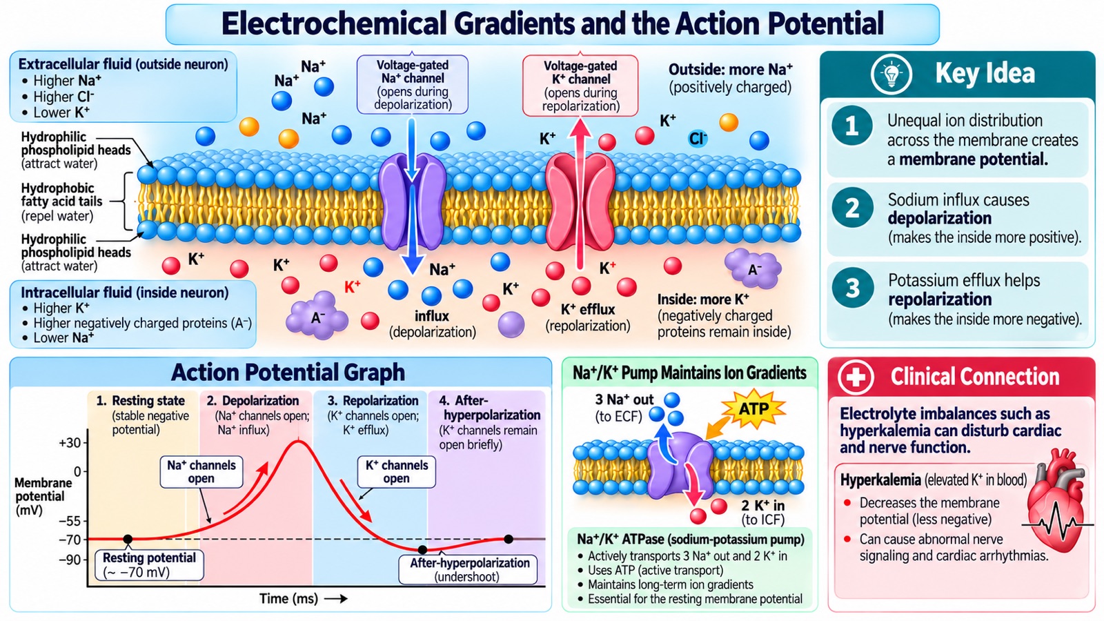
Figure 7. Selective ion movement produces electrical signals essential for nerve and muscle function.
Many human cells maintain electrical differences across their plasma membranes.
These differences are called membrane potentials.
6.1 Resting Membrane Potential
A typical resting neuron has a membrane potential near −70 mV.
This means the inside of the neuron is electrically negative relative to the outside.
The resting potential arises primarily from:
- Unequal ion distribution.
- Selective membrane permeability.
- Potassium permeability at rest.
- Ion gradients maintained by ATP-dependent transporters.
The Na⁺/K⁺ ATPase maintains long-term ion gradients and contributes a smaller direct electrogenic effect.
6.2 The Neuronal Action Potential
An action potential is a rapid change in membrane voltage that allows neurons to transmit electrical signals.
A typical neuronal action potential occurs through several stages.
Stage 1: Resting State
The neuron maintains a relatively negative membrane potential.
Stage 2: Threshold
A sufficient stimulus depolarizes the membrane to the threshold required to trigger a regenerative electrical response.
Stage 3: Depolarization
Voltage-gated sodium channels open.
Na⁺ enters the cell rapidly, making the inside more positive.
Stage 4: Repolarization
Sodium channels inactivate.
Outward potassium current increases, helping restore a more negative membrane potential.
Stage 5: Afterhyperpolarization
Potassium conductance may remain elevated briefly.
The membrane can become more negative than its usual resting potential.
Stage 6: Recovery
Ion channel states return toward baseline.
Membrane pumps maintain the concentration gradients necessary for repeated electrical signaling.
Important: The rapid rising and falling phases of an action potential are produced mainly by ion movement through channels, not by rapid cycling of the sodium–potassium pump.
6.3 Clinical Nursing Connection: Hyperkalemia
Potassium has a major influence on resting membrane potential.
When extracellular potassium becomes elevated, the potassium gradient changes.
This can alter cardiac cell excitability, sodium channel availability, and electrical conduction.
Severe hyperkalemia may cause dangerous cardiac dysrhythmias.
Possible ECG abnormalities include:
- Peaked T waves.
- PR interval prolongation.
- QRS widening.
- Severe conduction disturbances.
ECG changes are not always proportional to the severity of hyperkalemia.
Severe hyperkalemia requires urgent clinical evaluation and treatment.
7. Vesicular Transport: Moving Large Substances
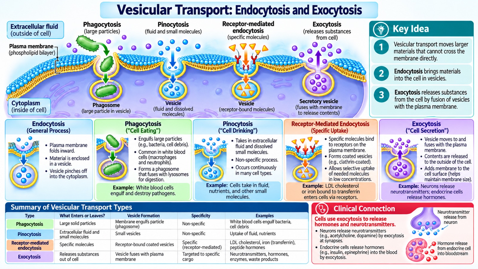
Figure 8. Vesicular transport allows cells to take up or release materials through membrane-bound vesicles.
Some substances are too large for conventional membrane channels or carrier proteins.
Cells can transport these substances through vesicles.
7.1 Endocytosis
Endocytosis brings material into a cell through membrane invagination and vesicle formation.
Several important forms exist.
Phagocytosis
Phagocytosis involves the engulfment of large particles.
Examples include:
- Bacteria.
- Cellular debris.
- Foreign particles.
Macrophages and neutrophils use phagocytosis as part of immune defense.
Pinocytosis
Pinocytosis involves the uptake of extracellular fluid and dissolved substances.
It occurs in many cell types.
Receptor-Mediated Endocytosis
Receptor-mediated endocytosis allows selective uptake of substances that bind to specific membrane receptors.
An example is cellular uptake of LDL cholesterol.
7.2 Exocytosis
Exocytosis releases substances when intracellular vesicles fuse with the plasma membrane.
Examples include:
- Neurotransmitter release.
- Insulin secretion.
- Other peptide hormone secretion.
- Release of certain enzymes and cellular products.
Clinical Nursing Connection
Vesicular transport plays essential roles in:
- Immune responses.
- Neurotransmission.
- Hormone regulation.
- Endocrine physiology.
- Cellular communication.
8. How Membrane Transport Maintains Whole-Body Homeostasis
Membrane transport mechanisms are not isolated events.
They support the functions of tissues, organs, and organ systems.
| Organ System | Important Membrane Processes | Homeostatic Function |
|---|---|---|
| Nervous | Sodium, potassium, calcium channels and ion pumps | Electrical signaling and neurotransmission |
| Cardiovascular | Sodium, potassium, calcium transport | Cardiac excitability and contraction |
| Respiratory | Oxygen and carbon dioxide diffusion | Gas exchange |
| Renal | Water channels, ion transporters, filtration | Fluid, electrolyte, and acid–base regulation |
| Gastrointestinal | Nutrient carriers and sodium-linked cotransport | Nutrient and fluid absorption |
| Endocrine | Hormone secretion and hormone-regulated transport | Metabolic and fluid regulation |
8.1 The Kidneys and Fluid Balance
The kidneys regulate the composition and volume of extracellular fluid.
Different nephron segments use specialized transport proteins to move:
- Sodium.
- Potassium.
- Chloride.
- Calcium.
- Hydrogen ions.
- Bicarbonate.
- Glucose.
- Water.
Hormones help adjust these processes.
Aldosterone promotes sodium reabsorption and potassium secretion in selected distal nephron segments.
Antidiuretic hormone (ADH), also called vasopressin, increases water permeability in the collecting ducts by regulating aquaporin insertion.
These mechanisms help regulate blood volume, blood pressure, electrolyte concentrations, and water balance.
8.2 The Heart and Electrical Stability
Cardiac cells depend on carefully regulated ion channels.
Sodium, potassium, and calcium movements determine important features of cardiac electrical activity.
Disruption of these processes can impair:
- Electrical conduction.
- Cardiac rhythm.
- Myocardial contraction.
- Overall circulation.
8.3 The Brain and Nervous System
Neurons rely on electrochemical gradients for electrical signaling.
Membrane channels also help regulate neurotransmitter release and cellular excitability.
Serious disturbances in oxygen delivery, glucose availability, or electrolyte balance can compromise neurological function.
9. Clinical Nursing Applications
Case 1: Hyponatremia and Brain-Cell Swelling
A patient develops a significantly reduced serum sodium concentration.
When extracellular fluid becomes sufficiently hypotonic, water may move into brain cells.
This can cause cerebral swelling.
Possible symptoms include:
- Headache.
- Nausea.
- Confusion.
- Seizures.
- Reduced consciousness.
Clinical Reasoning: The danger is not simply a low laboratory number. Reduced extracellular tonicity can alter brain-cell volume and function.
The severity, duration, and speed of onset are important.
Excessively rapid correction of chronic hyponatremia can also cause osmotic demyelination.
Treatment must follow appropriate clinical protocols.
Case 2: Hyperkalemia and Cardiac Dysrhythmias
A patient with impaired kidney function has a serum potassium concentration of 6.8 mEq/L.
Elevated extracellular potassium alters cardiac electrochemical gradients.
This may impair electrical conduction and increase the risk of severe dysrhythmias.
Clinical Reasoning: Potassium disorders affect the electrical behavior of cardiac cells.
This patient requires urgent assessment, appropriate monitoring, and treatment according to clinical protocols.
Case 3: Diarrhea and Dehydration
A patient experiences persistent diarrhea.
The patient may lose:
- Water.
- Sodium.
- Potassium.
- Bicarbonate.
These losses may cause dehydration, electrolyte abnormalities, and acid–base disturbances.
Appropriately formulated oral rehydration solution uses sodium–glucose cotransport to support intestinal water and electrolyte absorption.
Clinical Reasoning: Understanding the molecular transport mechanism explains why oral rehydration therapy works.
Case 4: Ischemia and Cellular Injury
A patient experiences an acute ischemic stroke.
Reduced blood flow limits the delivery of oxygen and metabolic substrates.
ATP production may decline.
As membrane transporters become less effective, ionic gradients deteriorate and intracellular water accumulation may occur.
Clinical Reasoning: Cellular dysfunction can begin with impaired energy metabolism and progress to tissue injury.
Restoring appropriate perfusion as rapidly and safely as clinically indicated is important for protecting vulnerable tissue.
10. NCLEX Review: Essential Associations
| Clinical or Physiological Finding | Important Mechanism |
|---|---|
| Oxygen entering cells | Simple diffusion |
| Carbon dioxide leaving cells | Simple diffusion |
| Glucose entering muscle through GLUT4 | Facilitated diffusion |
| Red blood cell swelling | Osmosis in a hypotonic environment |
| Red blood cell shrinking | Osmosis in a hypertonic environment |
| Three Na⁺ out and two K⁺ in | Primary active transport |
| Sodium and glucose entering intestinal epithelial cells together | Secondary active transport |
| Rapid neuronal depolarization | Voltage-gated sodium channels |
| Neuronal repolarization | Potassium efflux and sodium-channel inactivation |
| White blood cells engulfing bacteria | Phagocytosis |
| Neurotransmitter release | Exocytosis |
| Severe hyperkalemia | Altered cellular excitability and cardiac conduction |
11. NCLEX Practice Questions
Question 1
Which process allows oxygen to cross the plasma membrane without a specialized transport protein?
A. Primary active transport
B. Simple diffusion
C. Endocytosis
D. Sodium–glucose cotransport
Correct Answer: B. Simple diffusion
Rationale: Oxygen is a small, nonpolar molecule that can cross the phospholipid bilayer down its partial-pressure gradient.
Question 2
A red blood cell is placed in a strongly hypotonic solution. Which response should the nurse expect?
A. The cell shrinks.
B. The cell swells.
C. The cell immediately loses all intracellular potassium.
D. The cell remains unchanged.
Correct Answer: B. The cell swells.
Rationale: Water moves into the cell because the extracellular environment has a lower effective solute concentration.
Severe swelling can cause hemolysis.
Question 3
Which statement accurately describes the sodium–potassium ATPase?
A. It moves three sodium ions out and two potassium ions in per ATP hydrolyzed.
B. It moves two sodium ions out and three potassium ions in.
C. It transports equal amounts of sodium and potassium in both directions.
D. It transports three sodium ions into the cell and two potassium ions out.
Correct Answer: A.
Rationale: The Na⁺/K⁺ ATPase uses ATP to maintain sodium and potassium gradients by moving three Na⁺ ions outward and two K⁺ ions inward.
Question 4
Which mechanism is primarily responsible for the rapid rising phase of a typical neuronal action potential?
A. Sodium influx through voltage-gated channels
B. Sodium–potassium ATPase cycling
C. Water leaving the neuron
D. Exocytosis of proteins
Correct Answer: A. Sodium influx through voltage-gated channels
Rationale: Opening of voltage-gated sodium channels allows rapid sodium influx, causing membrane depolarization.
Question 5
Why does oral rehydration solution contain both glucose and sodium?
A. Glucose directly crosses the phospholipid bilayer without a transporter.
B. Sodium–glucose cotransport supports intestinal solute uptake and water absorption.
C. Water can enter cells only through primary active transport.
D. Potassium is the sole driver of glucose uptake.
Correct Answer: B.
Rationale: SGLT1 uses the sodium electrochemical gradient to transport glucose into intestinal epithelial cells, supporting sodium and water absorption.
12. Key Terminology
| Term | Definition |
|---|---|
| Plasma membrane | Selectively permeable boundary separating a cell from its environment |
| Phospholipid bilayer | Double layer of phospholipids forming the membrane's basic structure |
| Selective permeability | Ability of a membrane to permit some substances to cross more readily than others |
| Concentration gradient | Difference in a substance's concentration between regions |
| Electrical gradient | Difference in electrical potential |
| Electrochemical gradient | Combined chemical and electrical driving forces acting on an ion |
| Simple diffusion | Passive movement through the membrane without a transport protein |
| Facilitated diffusion | Passive movement through a channel or carrier protein |
| Osmosis | Net movement of water across a selectively permeable membrane |
| Tonicity | Effect of a solution on cell volume due mainly to nonpenetrating solutes |
| Active transport | Energy-dependent movement of substances against relevant gradients |
| ATPase | Enzyme that hydrolyzes ATP |
| Cotransport | Coupled movement of multiple substances through one transporter |
| Endocytosis | Uptake of extracellular material into membrane-bound vesicles |
| Exocytosis | Release of substances through fusion of vesicles with the plasma membrane |
| Membrane potential | Electrical potential difference across the cell membrane |
| Homeostasis | Regulation of internal conditions within physiologically compatible ranges |
Conclusion: Life Depends on Controlled Chemical Movement
The human body remains alive because its cells continually regulate what enters and leaves their membranes.
Every heartbeat, nerve impulse, muscle contraction, and metabolic reaction depends on maintaining appropriate chemical and electrical conditions.
Oxygen and nutrients must reach cells. Carbon dioxide and metabolic products must be transported away or processed. Water and electrolytes must remain balanced. Electrical gradients must be maintained so nerves and muscles can function.
The plasma membrane provides the selective barrier through which these essential processes occur.
Understanding membrane transport helps explain many clinical conditions, including:
- Electrolyte imbalances.
- Cardiac dysrhythmias.
- Dehydration.
- Edema.
- Diabetes mellitus.
- Kidney dysfunction.
- Neurological injury.
- Acid–base disorders.
The central lesson is simple: Life depends not only on which chemicals the body contains, but also on where those chemicals are located, how they move, and how carefully their concentrations are regulated.
Mastering cell membrane transport provides a foundation for understanding human physiology, pathophysiology, pharmacology, and clinical nursing.
Next Step
Membrane transport explains how chemicals move. The next question is what those chemicals actually are.
In upcoming articles, we will look more closely at the chemical level — atoms, ions, electrolytes, water, acids and bases, and pH — so that values like a serum potassium of 6.8 mEq/L mean something before they ever appear on a lab report.
References and Further Reading
- OpenStax. (2022). Anatomy and Physiology 2e — 3.1 The Cell Membrane. Rice University.
- OpenStax. (2022). Anatomy and Physiology 2e. Rice University.
- StatPearls. Physiology, Sodium Potassium Pump. NCBI Bookshelf.
Educational Disclaimer: This article is intended for nursing education and general scientific learning. Laboratory reference ranges, clinical assessments, and treatment approaches vary according to patient circumstances and institutional guidelines. The illustrations are original AI-generated educational graphics.