Understanding Bones: An Introduction to Bone Structure and Its 7 Functions
An illustrated nursing guide to what bones are made of, how they are built, and the seven jobs they do, from support and protection to blood cell production and hormone signaling.
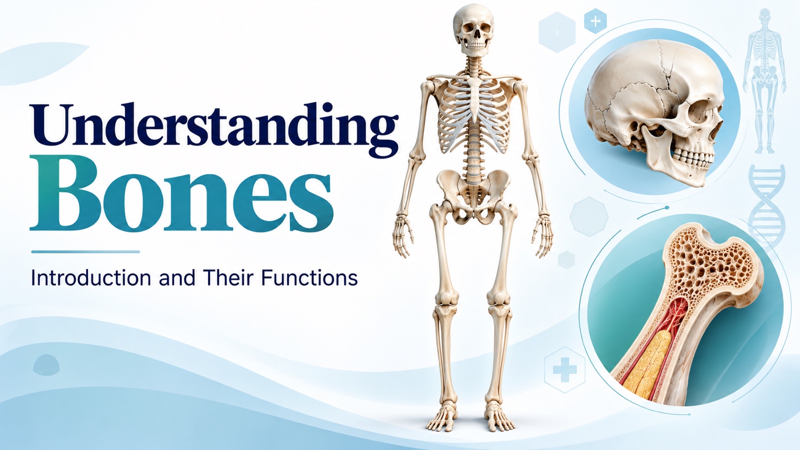
Most of us think of bones as the hard, dry framework we see hanging in a classroom skeleton.
That picture is misleading.
Living bone is a busy, blood-rich organ. It is constantly being broken down and rebuilt, it stores most of the body's calcium, it manufactures blood cells, and it even releases hormones that talk to the kidneys, pancreas, and muscles.
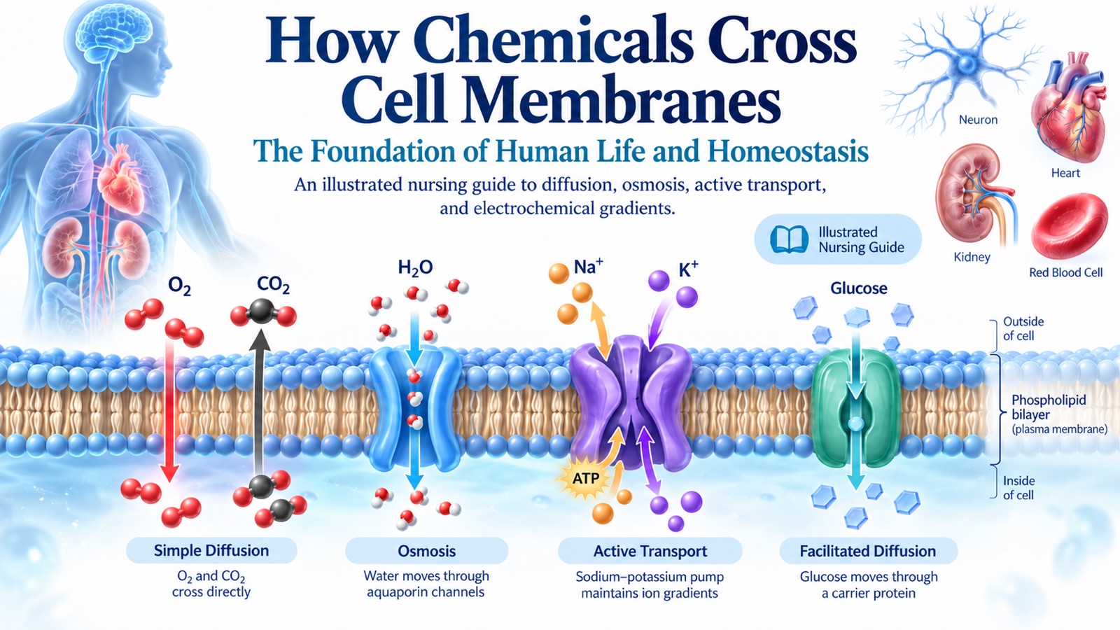
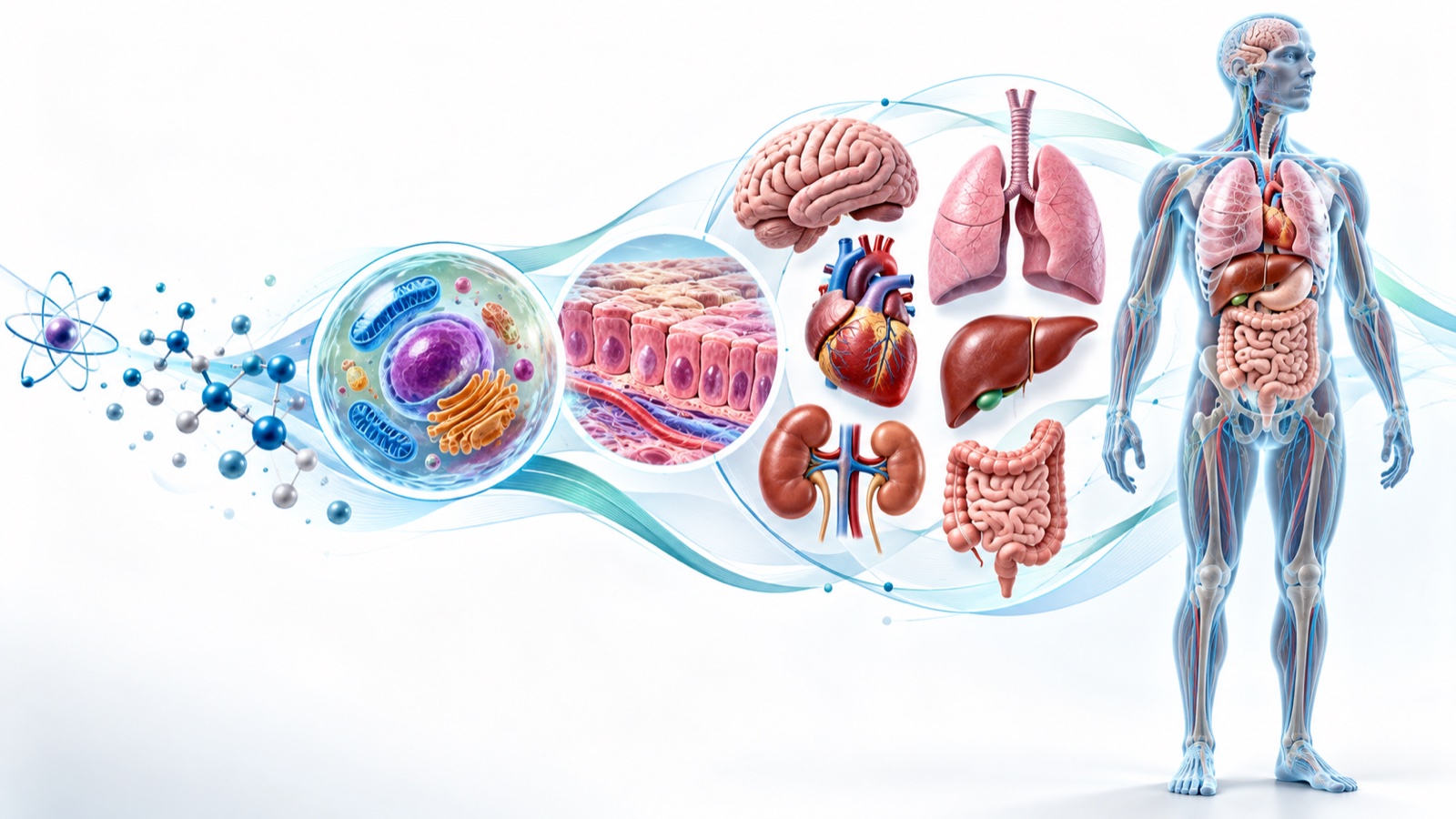
In From Atoms to Organism: The 8 Levels of Human Body Organization, we saw that organs are built from tissues working together. Bones are a perfect example. And in How Chemicals Cross Cell Membranes, we saw how tightly the body regulates electrolytes like calcium. Much of that calcium lives in bone.
This guide covers what bones are made of, how they are built, and the seven jobs they do every day — with the clinical connections nurses need to know.
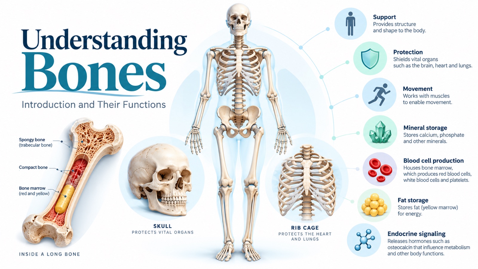
Figure 1. Bones do far more than hold the body up — they protect, move, store, produce, and signal.
Quick Overview: The 7 Functions of Bone
| Function | What Bones Do | Key Example |
|---|---|---|
| 1. Support | Provide the body's framework and bear weight | Spine, pelvis, and leg bones |
| 2. Protection | Shield vital organs from injury | Skull around the brain, ribs around the heart and lungs |
| 3. Movement | Act as levers that muscles pull on | Biceps flexing the elbow |
| 4. Mineral storage | Store and release calcium and phosphorus | About 99% of the body's calcium is in bone |
| 5. Blood cell production | Red marrow makes all blood cells | Red cells, white cells, and platelets |
| 6. Fat storage | Yellow marrow stores fat | Medullary cavity of adult long bones |
| 7. Endocrine regulation | Bone cells release hormones | Osteocalcin and FGF23 |
Introduction to Bones: What Bones Actually Are
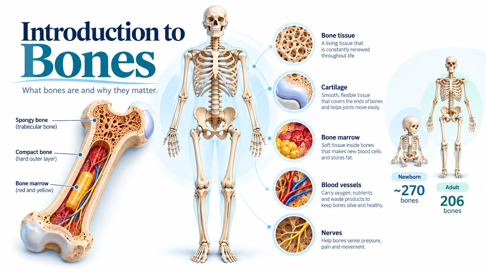
Figure 2. A bone is an organ made of several tissues: bone tissue, cartilage, marrow, blood vessels, and nerves.
Each bone is an organ, not just a lump of mineral.
A single bone contains:
- Bone (osseous) tissue — the hard, mineralized tissue that gives bone its strength.
- Cartilage — smooth, flexible tissue covering the ends of bones at joints.
- Bone marrow — soft tissue inside bones that makes blood cells and stores fat.
- Blood vessels — deliver oxygen and nutrients and remove waste.
- Nerves — allow bones to sense pressure and pain.
That last point explains something every nurse sees: fractures hurt intensely because the outer covering of bone is richly supplied with nerves.
How Many Bones Do We Have?
An adult skeleton has 206 bones.
A newborn has roughly 270 to 300 bony elements. Many of these fuse together during childhood and adolescence — for example, the separate plates of the infant skull and the five vertebrae that fuse to form the sacrum.
What Is Bone Made Of?
Bone matrix is a composite material with two main components:
| Component | Approximate Share | What It Contributes |
|---|---|---|
| Organic matrix (mostly type I collagen) | About one-third | Flexibility and resistance to twisting |
| Inorganic minerals (mainly calcium phosphate as hydroxyapatite) | About two-thirds | Hardness and resistance to compression |
Think of reinforced concrete: collagen is the steel rebar, and the mineral is the concrete.
Without enough mineral, bones become soft and bend, as in rickets or osteomalacia. Without healthy collagen, bones become brittle and break easily, as in osteogenesis imperfecta.
Compact Bone vs. Spongy Bone
Compact (cortical) bone is the dense outer layer. It makes up roughly 80% of skeletal mass and is organized into tightly packed cylindrical units called osteons.
Spongy (trabecular or cancellous) bone is found inside bones, especially at their ends. It is a lattice of thin struts called trabeculae, which are lined up along lines of stress. The spaces between them hold red bone marrow.
Spongy bone is lighter but has a much larger surface area, so it is more metabolically active — and it is often the first to lose density in osteoporosis.
The Anatomy of a Long Bone
Long bones such as the femur and humerus have a standard layout:
- Diaphysis: The long shaft, made mostly of compact bone.
- Epiphyses: The expanded ends, filled with spongy bone.
- Metaphysis: The region where the shaft widens toward each end.
- Epiphyseal plate (growth plate): A layer of cartilage that allows bones to lengthen during childhood. After growth ends, it ossifies into the epiphyseal line.
- Medullary cavity: The hollow center of the shaft, containing marrow.
- Periosteum: A tough membrane covering the outside of the bone, rich in nerves and blood vessels.
- Endosteum: A thin membrane lining the inner surfaces.
- Articular cartilage: Smooth hyaline cartilage on the joint surfaces.
Nursing note: Growth-plate injuries in children need careful assessment because damage can affect future bone length.
Bone Cells: The Builders, Maintainers, and Demolition Crew
| Cell | Role | Memory Tip |
|---|---|---|
| Osteogenic cells | Stem cells that become osteoblasts | The reserves |
| Osteoblasts | Build new bone matrix | OsteoBlasts Build |
| Osteocytes | Mature bone cells that maintain matrix and sense mechanical load | The supervisors |
| Osteoclasts | Break down (resorb) bone and release minerals | OsteoClasts Carve |
These cells work together in a process called bone remodeling.
Old or damaged bone is removed by osteoclasts and replaced by osteoblasts. In adults, roughly 10% of the skeleton is remodeled each year.
This is why bones respond to exercise, heal after fractures, and lose density during prolonged bed rest.
Bones Come in Different Shapes
| Shape | Description | Examples |
|---|---|---|
| Long | Longer than they are wide | Femur, humerus, phalanges |
| Short | Roughly cube-shaped | Carpals (wrist), tarsals (ankle) |
| Flat | Thin and often curved | Skull bones, sternum, ribs, scapula |
| Irregular | Complex shapes | Vertebrae, many facial bones |
| Sesamoid | Small bones embedded in tendons | Patella (kneecap) |
Shape usually reflects function. Long bones act as levers, flat bones protect, and short bones allow stable but flexible movement.
Function 1: Support
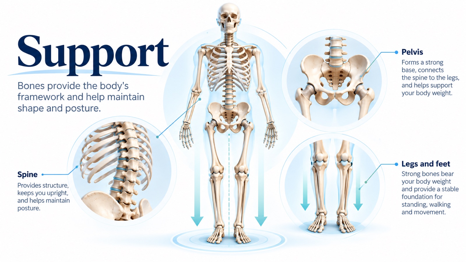
Figure 3. The skeleton provides a framework that supports body weight and maintains posture.
The skeleton is the body's internal scaffolding.
Without it, soft tissues would have nothing to attach to and no way to hold their shape.
- The spine keeps the body upright, carries the weight of the head and trunk, and maintains posture through its natural curves.
- The pelvis forms a strong base that transfers weight from the spine to the legs.
- The leg and foot bones bear body weight and provide a stable foundation for standing and walking.
Bones also adapt to the loads placed on them. Weight-bearing activity stimulates bone formation, while lack of loading leads to bone loss.
Clinical Nursing Connection
Immobility is hard on the skeleton.
Patients on prolonged bed rest, and astronauts in microgravity, lose bone mass quickly. Calcium released from bone can raise urinary calcium and increase the risk of kidney stones.
Early mobilization, weight-bearing as permitted, and fall-prevention strategies all help protect the skeleton.
Function 2: Protection
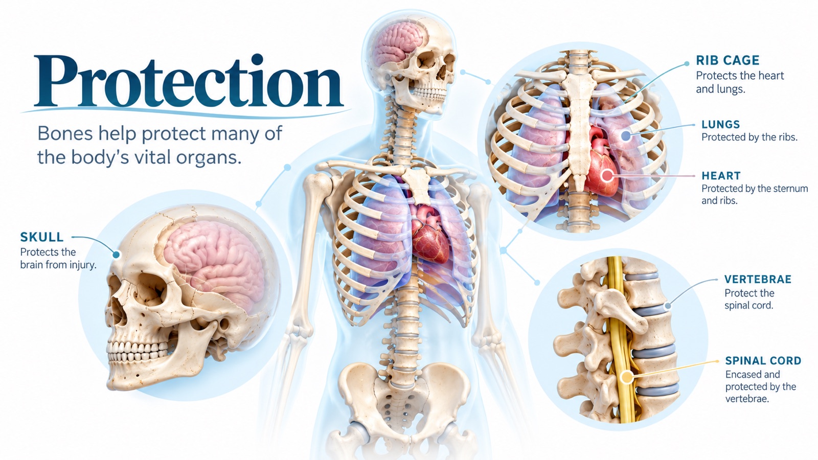
Figure 4. Bones form protective enclosures around the brain, heart, lungs, and spinal cord.
Some of the body's most vulnerable organs are surrounded by bone.
| Bone Structure | What It Protects |
|---|---|
| Skull (cranium) | Brain |
| Rib cage and sternum | Heart and lungs |
| Vertebral column | Spinal cord |
| Pelvis | Bladder, reproductive organs, and part of the intestines |
The rib cage is a clever design. It protects the heart and lungs but stays flexible enough to expand with every breath.
Clinical Nursing Connection
When protective bones break, the injury beneath them is often the bigger concern:
- Skull fractures raise concern for traumatic brain injury. Monitor level of consciousness, pupils, and signs of increased intracranial pressure.
- Rib fractures can injure the lungs and cause pneumothorax. Pain also leads patients to breathe shallowly, which increases the risk of atelectasis and pneumonia — so pain control and deep breathing are key nursing priorities.
- Vertebral fractures can threaten the spinal cord. Spinal precautions remain in place until injury has been ruled out.
Function 3: Movement
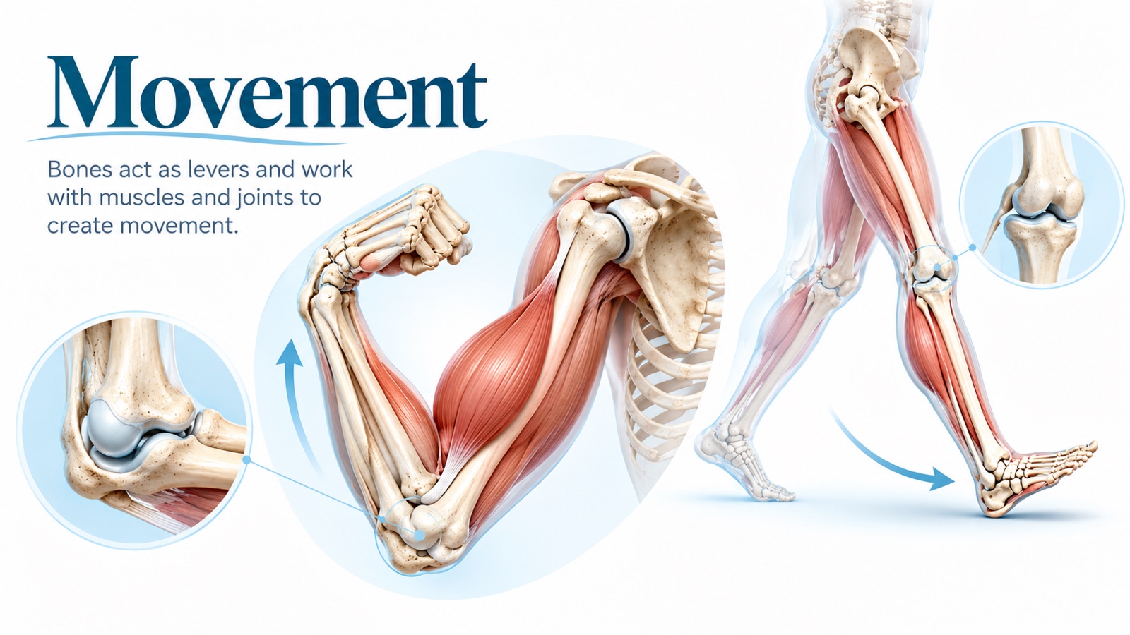
Figure 5. Bones act as levers, joints act as pivot points, and muscles provide the pulling force.
Bones cannot move on their own.
Movement happens when three structures work together:
- Bones act as rigid levers.
- Joints act as pivot points (fulcrums).
- Muscles contract and pull on bones through tendons.
For example, when the biceps contracts, it pulls on the radius and flexes the elbow. When the calf muscles contract, they lift the heel and push the body forward during walking.
Ligaments connect bone to bone and stabilize joints.
Quick distinction: Tendons connect muscle to bone. Ligaments connect bone to bone.
Clinical Nursing Connection
Joint and musculoskeletal problems — arthritis, fractures, joint replacements, and contractures — directly limit movement.
Nurses assess range of motion, gait, and mobility, and help prevent complications such as contractures and pressure injuries in patients who cannot move independently.
Function 4: Mineral Storage and Release
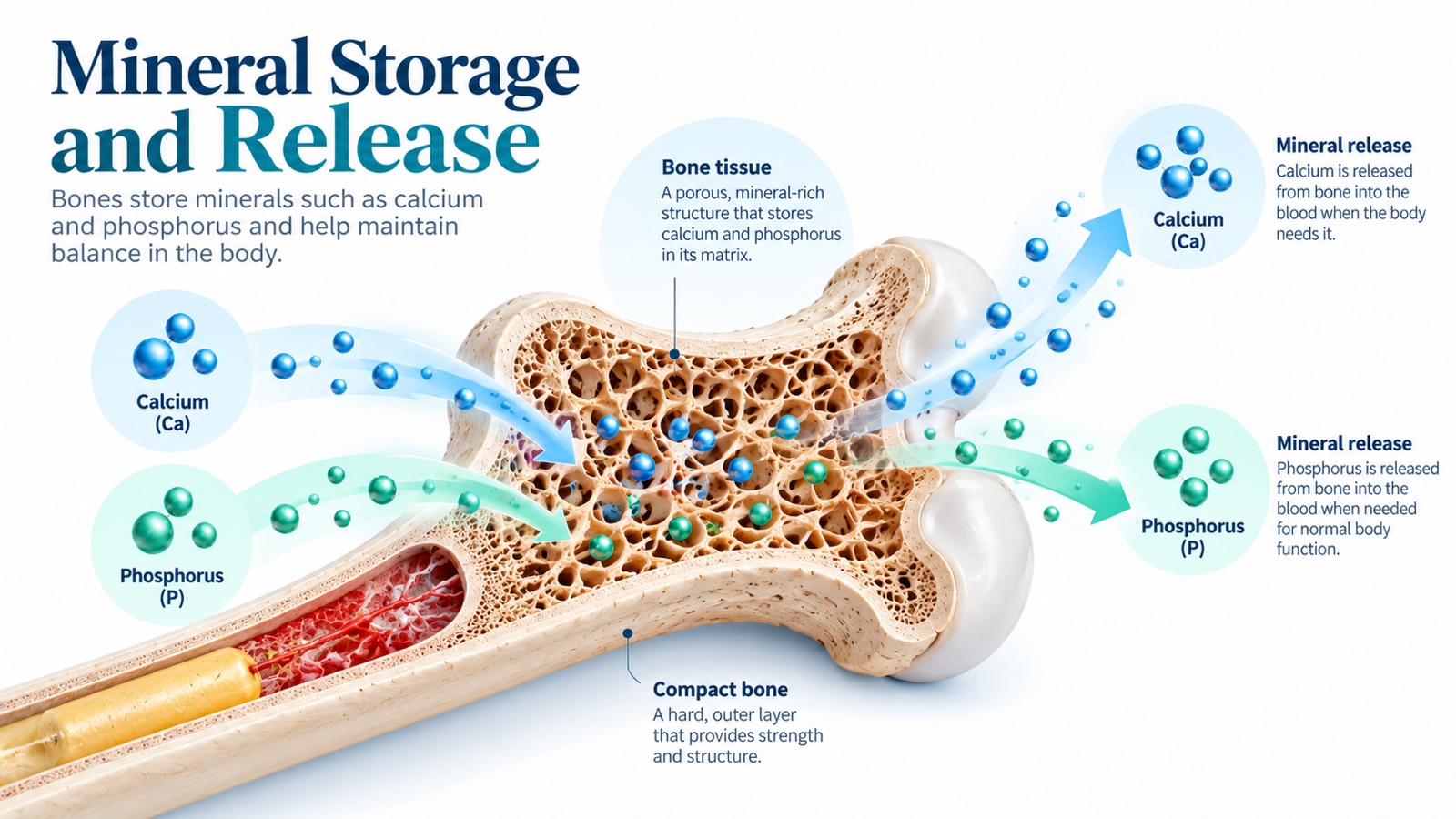
Figure 6. Bone acts as a mineral reservoir, storing calcium and phosphorus and releasing them into the blood as needed.
Bone is the body's mineral bank.
It holds about 99% of the body's calcium and roughly 85% of its phosphorus.
These minerals are not locked away permanently. Bone constantly deposits and withdraws them to keep blood levels within a narrow range.
That matters because calcium in the blood is essential for:
- Muscle contraction, including the heart.
- Nerve signaling.
- Blood clotting.
- Hormone and neurotransmitter release.
How the Body Controls Blood Calcium
| Hormone | Released When | Main Effects |
|---|---|---|
| Parathyroid hormone (PTH) | Blood calcium falls | Increases osteoclast activity to release calcium from bone, increases kidney calcium reabsorption, and activates vitamin D |
| Calcitriol (active vitamin D) | Stimulated by PTH | Increases calcium and phosphate absorption from the intestine |
| Calcitonin | Blood calcium rises | Inhibits osteoclasts; has a relatively modest role in adults |
In other words, when blood calcium drops, the body will borrow from bone to protect the heart and nerves.
If that borrowing continues for years — because of low dietary calcium, vitamin D deficiency, or hormonal changes — bone density gradually declines.
Clinical Nursing Connection
- Osteoporosis: Loss of bone mass and microarchitecture increases fracture risk, especially of the hip, spine, and wrist. Calcium, vitamin D, weight-bearing exercise, fall prevention, and medication adherence are central to nursing education.
- Hypercalcemia: Cancer that has spread to bone can release large amounts of calcium into the blood, causing fatigue, constipation, confusion, kidney stones, and cardiac rhythm changes.
- Hypocalcemia: Low calcium increases neuromuscular excitability, which can cause tingling, muscle cramps, tetany, and seizures.
A typical adult total serum calcium reference range is about 8.5–10.5 mg/dL, although ranges vary by laboratory.
Function 5: Blood Cell Production
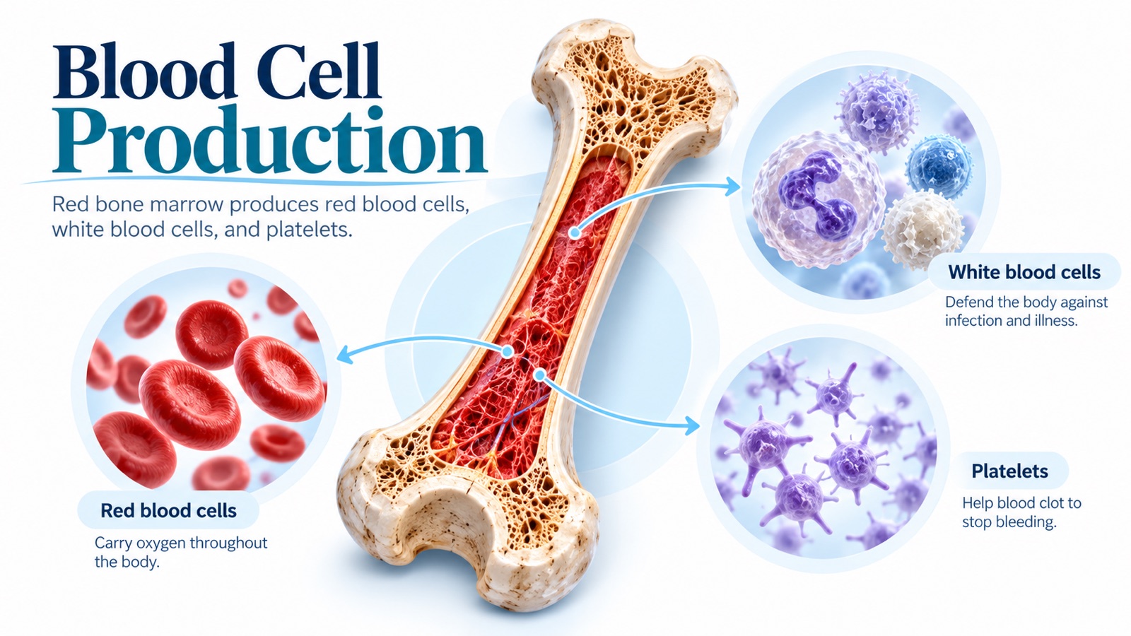
Figure 7. Red bone marrow produces red blood cells, white blood cells, and platelets.
Hematopoiesis is the production of blood cells, and in healthy adults it takes place in red bone marrow.
Red marrow produces:
| Cell Type | Main Job |
|---|---|
| Red blood cells (erythrocytes) | Carry oxygen using hemoglobin |
| White blood cells (leukocytes) | Defend the body against infection |
| Platelets (thrombocytes) | Help blood clot and stop bleeding |
The scale is remarkable: the marrow produces roughly 2 million red blood cells every second.
In infants, red marrow fills most bones. In adults, it is concentrated mainly in the flat and irregular bones — the skull, sternum, ribs, vertebrae, and pelvis — and the upper ends of the femur and humerus.
Clinical Nursing Connection
- Bone marrow suppression: Chemotherapy, radiation, and some medications can reduce marrow output. This can cause anemia (fatigue, shortness of breath), neutropenia (high infection risk), and thrombocytopenia (bleeding risk). Nurses monitor blood counts and implement infection and bleeding precautions.
- Bone marrow aspiration and biopsy: Usually performed at the posterior iliac crest, because the pelvis contains active red marrow.
- Intraosseous (IO) access: In emergencies, fluids and medications can be given directly into the marrow cavity, commonly at the proximal tibia or proximal humerus, because marrow drains into the circulation.
Function 6: Fat Storage
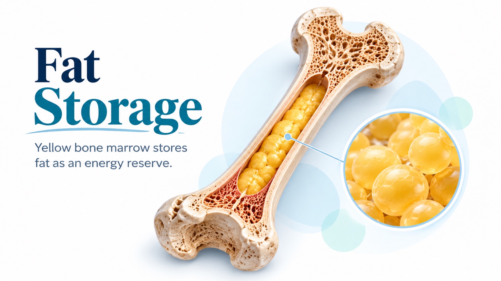
Figure 8. Yellow bone marrow in the medullary cavity stores fat as an energy reserve.
As we age, much of the red marrow in the shafts of long bones is gradually replaced by yellow bone marrow.
Yellow marrow is made mostly of fat cells (adipocytes). It serves as an energy reserve.
It is not completely inactive, though. In situations such as severe blood loss or chronic anemia, yellow marrow can convert back into red marrow to increase blood cell production.
Clinical Nursing Connection: Fat Embolism
After a fracture of a long bone such as the femur — or after orthopedic surgery — fat droplets from the marrow can enter the bloodstream.
Fat embolism syndrome typically appears within about 24–72 hours and may cause:
- Sudden shortness of breath and low oxygen saturation.
- Confusion or other neurological changes.
- A petechial rash, often on the chest, axillae, or conjunctiva.
Prompt recognition and early stabilization of long-bone fractures are important. Report respiratory or mental status changes immediately.
Function 7: Endocrine Regulation
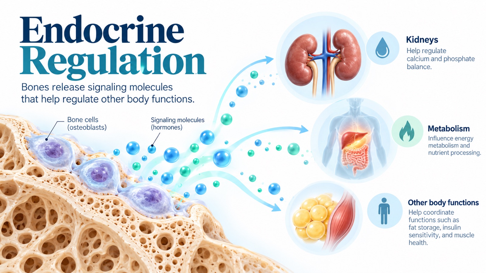
Figure 9. Bone cells release hormones that influence the kidneys, metabolism, and other tissues.
This is the newest and most surprising function of bone.
Bone is not only a target of hormones — it also produces them.
| Hormone | Made By | Main Effects |
|---|---|---|
| FGF23 (fibroblast growth factor 23) | Osteocytes and osteoblasts | Signals the kidneys to excrete phosphate and reduces activation of vitamin D |
| Osteocalcin | Osteoblasts | Linked to insulin secretion and sensitivity, energy metabolism, and muscle function |
FGF23 is well established. It helps explain why patients with chronic kidney disease develop disorders of phosphate, calcium, and bone together — a condition called CKD–mineral and bone disorder.
Osteocalcin's metabolic effects are strongly supported in animal studies, while research in humans is still evolving.
Key Concept: Bone is part of a body-wide communication network. It exchanges signals with the kidneys, gut, parathyroid glands, pancreas, fat, and muscle.
Nursing Connection: Why Bone Health Matters
Bone problems rarely stay limited to the skeleton.
Consider an older adult who falls and fractures a hip:
- Years of gradual bone loss weaken the femoral neck.
- A fall causes a fracture — often with a shortened, externally rotated leg.
- Pain and surgery limit mobility.
- Immobility increases the risk of blood clots, pneumonia, pressure injuries, and delirium.
- Recovery may be prolonged, and independence may be lost.
A single weakened bone can become a whole-body problem — the same pattern we saw in the levels of body organization.
NCLEX Practice Questions
Question 1
A patient receiving chemotherapy has a platelet count well below normal. Which nursing action is most appropriate?
A. Encourage the use of a straight razor
B. Implement bleeding precautions
C. Administer intramuscular injections as ordered without changes
D. Encourage vigorous contact sports
Correct Answer: B. Implement bleeding precautions
Rationale: Chemotherapy can suppress red bone marrow, reducing platelet production. Low platelets increase bleeding risk, so the nurse should use measures such as soft toothbrushes, electric razors, and avoiding unnecessary injections.
Question 2
Two days after a femur fracture, a patient suddenly becomes confused and short of breath, and petechiae appear on the chest. What should the nurse suspect?
A. Fat embolism syndrome
B. Hypocalcemia
C. Compartment syndrome of the arm
D. Osteoporosis
Correct Answer: A. Fat embolism syndrome
Rationale: Fat from the yellow marrow of long bones can enter the circulation after a fracture. The classic pattern includes respiratory distress, neurological changes, and a petechial rash.
Question 3
Which bone cell is primarily responsible for breaking down bone and releasing calcium into the blood?
A. Osteoblast
B. Osteocyte
C. Osteoclast
D. Chondrocyte
Correct Answer: C. Osteoclast
Rationale: Osteoclasts resorb bone matrix. Parathyroid hormone increases their activity when blood calcium falls.
Question 4
A patient has three fractured ribs. Which nursing intervention best prevents a respiratory complication?
A. Keep the patient flat and still
B. Restrict fluids
C. Provide adequate pain control and encourage deep breathing
D. Discourage coughing at all times
Correct Answer: C. Provide adequate pain control and encourage deep breathing
Rationale: Pain from rib fractures causes shallow breathing, which increases the risk of atelectasis and pneumonia. Effective pain management allows the patient to breathe deeply and clear secretions.
Final Summary
| Function | Main Idea |
|---|---|
| Support | Bones form the body's framework and bear its weight |
| Protection | Bones shield the brain, heart, lungs, and spinal cord |
| Movement | Bones act as levers for muscles, pivoting at joints |
| Mineral storage | Bone stores calcium and phosphorus and releases them as needed |
| Blood cell production | Red marrow produces red cells, white cells, and platelets |
| Fat storage | Yellow marrow stores fat and can revert to red marrow if needed |
| Endocrine regulation | Bone cells release hormones such as FGF23 and osteocalcin |
Bones are living organs that are constantly being rebuilt.
They hold us up, protect us, move us, feed our blood, and help regulate the chemistry of the entire body.
When a nurse assesses a fracture, a calcium level, or a blood count, they are really assessing the health of the skeleton.
Next Step
Now that we know what bones do, the next step is to look at how they connect and move.
In upcoming articles, we will explore joints, cartilage, and the muscular system — the structures that turn a rigid skeleton into a body that can walk, lift, and breathe.
References and Further Reading
- OpenStax. (2022). Anatomy and Physiology 2e — 6.1 The Functions of the Skeletal System. Rice University.
- OpenStax. (2022). Anatomy and Physiology 2e — 6.3 Bone Structure. Rice University.
- OpenStax. (2022). Anatomy and Physiology 2e — 6.7 Calcium Homeostasis: Interactions of the Skeletal System and Other Organ Systems. Rice University.
- National Institute of Arthritis and Musculoskeletal and Skin Diseases. Osteoporosis. National Institutes of Health.
Educational Disclaimer: This article is intended for nursing education and general scientific learning. Laboratory reference ranges, clinical assessments, and treatment approaches vary according to patient circumstances and institutional guidelines. The illustrations are original AI-generated educational graphics.